The Value of Acoustic Reflexes in Practice: A Retrospective Look

From time to time there is invariably a question about the utility of obtaining acoustic reflexes. This stapedius muscle contraction to intense sound levels measured at ear level can provide important diagnostic information to the audiologists. However because of many variables that can impact its measurement, obtaining them can be a source of frustration for the audiologist. Questions often arise, such as “Do we really need to do them?” or more often than not, “If we have to do them, are ipsilateral reflexes adequate?”
Survey evidence from the US suggests that acoustic reflex measurements are conducted by the large majority of audiologists, but only about two-thirds (65%) include them as part of the standard test battery.1 Moreover, only 60% report measurement of ipsilateral and contralateral reflexes, with a third collecting only ipsilateral measures and a much smaller proportion (6%) collecting only contralateral measures. This is unfortunate. The notion that hearing loss is usually confined to the inner or middle ear has been clearly dispelled by findings showing noise- and age-induced auditory nerve dysfunction,2,3 and the acoustic reflex remains the only probe of central auditory function typically included in the standard test battery. Neglecting this simple assessment of central auditory function compromises our ability to detect retrocochlear dysfunction and diminishes the value of associated diagnoses.
Much of what we know currently about the human acoustic reflex arc is based on the work of Borg on rabbits4 with considerable contributions from Møller.5 The reflex is mediated by the stapedius muscle in the middle ear, which contracts in response to moderate-to-loud acoustic stimulation, reducing the admittance of the ossicular chain. The lowest level of acoustic energy needed to produce a significant change in admittance, generally defined as at least 0.02 mmho, is called the acoustic reflex threshold. The reflex is consensual, so that when one ear is stimulated, both pathways respond. Therefore there are two recording conditions possible: an ipsilateral condition where the stimulus is presented to the same ear in which the measurement probe is monitoring immittance, and a contralateral condition where the sound is presented contralaterally to the ear in which immittance is being monitored. The general practice is use pure tone stimuli at 500, 1000 and 2000 Hz, although 4000 Hz is sometimes also used. There may be times when broadband noise is used as the stimulus, which can be compared to pure tone reflex data to provide objective information about the status of the cochlea (see Hall & Swanepoel6 for information on the Sensitivity Prediction by Acoustic Reflex or SPAR). Normative data for acoustic reflex thresholds are available for normal hearing and for varying degrees of cochlear hearing loss, and range from 75 to 90 dB HL. Generally the intensity needed to elicit a response increases with increasing levels of cochlear hearing loss.
The reflex is triggered when an ear is stimulated with a moderately intense signal that reaches the ventral cochlear nucleus (CN) and is projected via one of two tracts: one leading directly to the facial nerve nucleus on the same side and another that crosses the midline of the brainstem and connects with the contralateral superior olivary complex (SOC). The cochlear nucleus also sends a tract to the ipsilateral SOC which connects to its partner in the contralateral field, as well as both facial nerve nuclei, both ipsilaterally and contralaterally. The efferent portion of the pathway from the facial nerve nucleus to the facial nerve runs through the internal auditory meatus and into the middle ear where it connects with the stapedius muscle.7 Because of the complexity of the arc and its involvement with both peripheral and central mechanisms, it can be impacted by outer, middle, and inner ear pathologies, as well as by pathology of the auditory nerve and lower brainstem. Reflexes are typically not present for conductive hearing losses.
One of the most common methods to characterize different patterns of acoustic reflex activation in relation to site of lesion was developed by Jerger8,9 (see Hall and Swanepoel6 for further information), and includes five patterns based on both ipsilateral and contralateral measures: “vertical” (reflexes absent in one probe/measurement ear), “diagonal” (reflexes absent in one stimulus ear), “horizontal” (both contralateral reflexes absent), “inverted L” (both contralateral reflexes absent and one unilateral reflex absent) and “uni-box” (one reflex absent). Along with audiometric and tympanometric results, acoustic reflex recordings that typify these five patterns can be used to provide an initial estimate of the site of lesion.
Acoustic reflexes are remarkably sensitive to retrocochlear dysfunction given the ease with which they can be obtained.10,11 In a study of 30 individuals with surgically confirmed cerebellopontine angle tumours and 30 controls matched for hearing loss, the crossed acoustic reflex was found to have a sensitivity of 83% and a specificity of 97%.12 Similarly, a study of 89 individuals with VIIIth nerve tumours found the sensitivity of the reflex to be 84%.13 Reflexes are also sensitive to auditory neuropathy: In a large multi-site study, acoustic reflexes were either absent or elevated in all individuals with auditory neuropathy that were subjected to the measurement (n = 148).14
Suspicion of neural dysfunction clearly warrants electrophysiological assessment and a referral for magnetic resonance imaging, but if reflexes are not measured in the first place, there may be no grounds for suspicion. Recall that significant threshold asymmetries are only present in about 80–90% of individuals with confirmed lesions (depending on the criteria selected for a identifying a significant asymmetry)15 and abnormal reflexes can be found in cases of retrocochlear dysfunction even when thresholds are normal. Abnormal speech scores are present in a still smaller proportion of cases with retrocochlear involvement.10
In a retrospective review of clients at the Nova Scotia Hearing and Speech Centres with confirmed lesions of the auditory nerve or low brainstem, the acoustic reflex data tell an interesting tale. Of the 34 clients (aged from 10 to 64 years), with thresholds ranging from normal limits to moderate sensorineural hearing loss, 88% had abnormal acoustic reflex patterns following Jerger’s patterns as noted above (see Figure 1). However, if one chooses to focus on the subset of clients with clinically normal hearing (see Table 1), the value of the acoustic reflex is startlingly obvious. In ALL of the cases of confirmed lesions in the presence of normal hearing (and excellent high-intensity word recognition scores), the acoustic reflex pattern was abnormal. Stated differently, if the audiologist had chosen to perform only pure tone threshold testing and high intensity word recognition (with a presentation level of 80 or 90 dB HL) and omit the acoustic reflex test from the audiological battery, these lesions would have be missed.
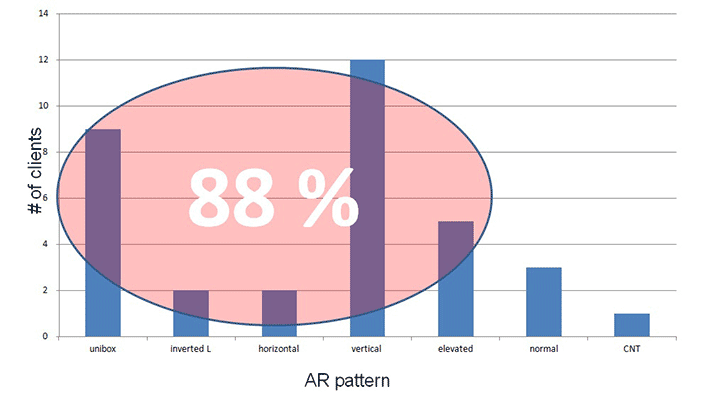
Figure 1. Acoustic reflex pattern data from a retrospective study of 34 clients with confirmed retrocochlear pathology.
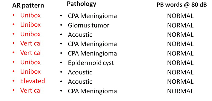
Table 1. Acoustic Reflex Data from Nine Clients with Clinically Normal Hearing and Confirmed Retrocochlear Pathology.
As professionals who are often the first point of contact for individuals experiencing hearing difficulties, it is critical that our assessments are comprehensive enough to guide treatment and to indicate appropriate medical referrals. This means that we should not be willing to forgo assessment of central auditory function. The acoustic reflex, with its proven track record for detecting peripheral and central dysfunction when measured ipsilaterally and contralaterally, and its simple procedural requirements, should be part of every audiologist’s standard test battery.
References
- Emanuel DC, Henson OE, Knapp RR. Survey of audiological immittance practices. Am J Audiol 2012;21:60–75.
- Gates GA, Feeney MP, Higdon RJ. Word recognition and the articulation index in older listeners with probable age-related auditory neuropathy. J Am Acad Audiol 2003;14:574–81.
- Kujawa SG, Liberman MC. Adding insult to injury: cochlear nerve degeneration after "temporary" noise-induced hearing loss. J Neurosci 2009;29:14077–85.
- Borg E. On the neuronal organization of the acoustic middle ear reflex: A physiological and anatomical study. Brain Res 1973;49:101–23.
- Møller AR. Hearing; Anatomy, Physiology, and Disorders of the Auditory System. San Diego: Plural Publishing; 2012.
- Hall JW, Swanepoel deW. Objective Assessment of Hearing. San Diego: Plural Publishing; 2010.
- Musiek F, Baran J. The Auditory System: Anatomy, Physiology and Clinical Correlates. Boston: Pearson, Allyn & Bacon; 2007.
- Jerger S, Jerger J. Diagnostic value of crossed versus uncrossed acoustic reflexes: Eighth nerve and brainstem disorders. Arch Otolaryngol 1977;103:445–53.
- Jerger J, Jerger S, Hall JW III. A new acoustic reflex pattern. Arch Otolaryngol 1979;105:24–28.
- Bergenius J, Borg E, Hirsch A. Stapedius reflex test, brainstem audiometry and opto-vestibular tests in diagnosis of acoustic neurinomas. A comparison of test sensitivity in patients with moderate hearing loss. Scand Audiol 1983;12:3–9.
- Hirsch A, Anderson H. Audiologic test results in 96 patients with tumours affecting the eighth nerve. A clinical study with emphasis on the early audiological diagnosis. Acta Oto-laryngol Suppl 1980;369:1–26.
- Olsen WO, Bauch CD, Harner SG. Application of Silman and Gelfand (1981) 90th percentile levels for acoustic reflex thresholds. J Speech Hear Dis 1983;48:330–32.
- Godey B, Morandi X, Beust L, et al. Sensitivity of auditory brainstem response in acoustic neuroma screening. Acta Otolaryngol;118:501–504.
- Berlin CI, Hood LJ, Morlet T, et al. Multi-site diagnosis and management of 260 patients with auditory neuropathy/dys-synchrony (auditory neuropathy spectrum disorder). Int J Audiol 2010;49:30–43.
- Cheng TC, Wareing MJ. Three-year ear, nose, and throat cross-sectional analysis of audiometric protocols for magnetic resonance imaging screening of acoustic tumors. Otolaryngol Head Neck Surg 2012;146:438–447.