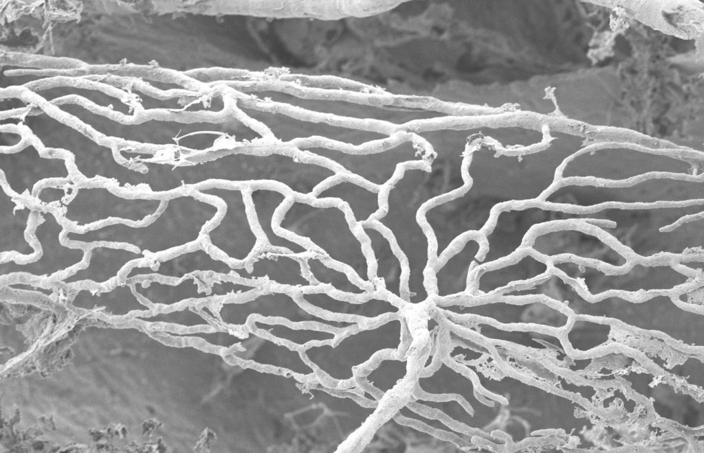
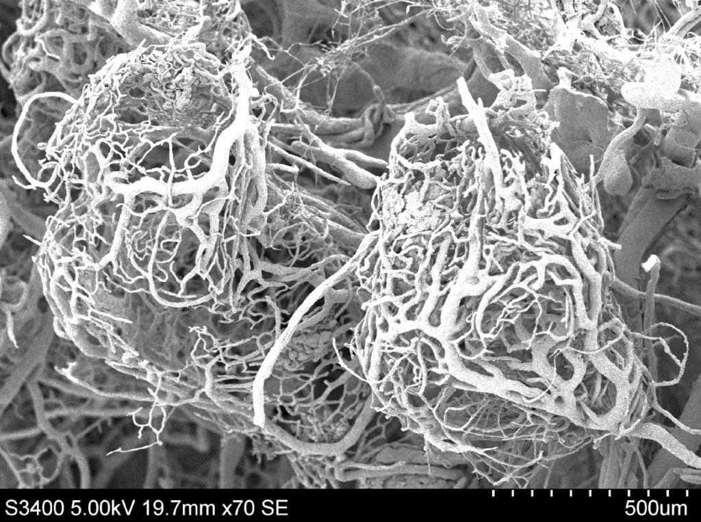
What Are These (Unusual) Images of Inner Ear Structures? A Picture Quiz.
From the Labs to the Clinics
Renowned auditory researcher Dr. Robert Harrison brings us up to date on information and research from the Labs. Appropriately titled “From the Labs to the Clinics”, Bob is involved in laboratory and applied/clinical research, including evoked potential and otoacoustic emission studies and behavioural studies of speech and language development in children with cochlear implants. For a little insight into Bob’s interests outside the lab and the clinic, we invite you to climb aboard Bob’s Garden Railway.
I have recently started to sort through hundreds of photo-micrographs accumulated during my decades of research studies. I have images taken with the light microscope (of various types) and images from electron-microscopes. Typically, in an anatomical study, the final paper will contain a few good representative images selected from the large pile of micrographs taken. However, I have come across interesting images that have not been central to the research studies and are not included in publications but are nevertheless interesting. Whilst I have ownership of these images, they were really shot by team members in my lab. For the images shown here, I want to credit lab technicians Richard Mount and Jaina Negandhi, and graduate trainee Mattia Carraro (PhD awarded in 2016).
Most of my research has been on the inner ear, so I’ve picked out three unusual images that, as audiologists, you might recognize or not. Perhaps we can make this a mystery game where you first look at the picture and try to identify the structure before reading my detailed description.


I trust that you were close to being correct in identifying these images. Look out for more mystery picture quizzes in future issues of Canadian Audiologist.

