
Sensory Neural Asymmetry Associated with a Long-Standing Conductive Component – A Case Study


Introduction
This is the case of a 67-year-old male who was a drummer since age 5. He had noticed left hearing loss for several years but denied any problems with the right ear. This case presentation raises the question of whether there can be protection from loud noise and music in the presence of a long-standing conductive hearing loss.
There is a long history of literature dating back almost a half century in which Schuknecht and his colleagues (see, for example, Walby, Barrera, and Schuknecht, 1983) showed that toxic molecules can permeate across the round window, causing sensorineural pathology as a secondary sequela of chronic otitis media. Schachern et al. (1987) showed that the round window was semipermeable to toxins and other “larger” molecules, which can result in a sensorineural component with long-lasting otitis media. And in a study of shipyard workers in Ulsan, Korea, they found hearing threshold changes to be lower in the ear with conductive hearing loss than in the opposite ear; this effect was greatest at 4000 Hz (Park et al., 2016). However, in 1991, a retrospective study in the United Kingdom concluded that there were no significant sensory neural differences in hearing thresholds between ears with conductive loss and those without (Simpson et al., 1991). More longitudinal studies with younger subjects are needed.
There are, however, fewer studies showing that long-standing conductive hearing loss can act as a “permanent hearing protector” of the cochlea.
Studies have been inconsistent regarding whether conductive hearing loss protects the inner ear from noise. This can vary depending on the type of conductive loss. Note that chronic middle ear dysfunction could theoretically provide more protection than stapes fixation in otosclerosis. Stapes fixation essentially destroys the stapedius muscle reflex (Forli et al., 2025). The stapedius muscle reflex is known to attenuate loud sounds to a minimal extent, primarily at low frequencies (Zakrisson, 1975). This occurs by inducing stiffness of the ossicles in the middle ear (Trevino et al., 2023). The magnitude of protection from this reflex varies depending on the type of exposure: impulse noise or steady-state noise. An example of impulse noise in this patient’s history is his own drums, while a steady-state noise example may be a factory machine running continuously for hours at the same sound level. The stapedial reflex adapts quickly, causing the muscle to relax after 10 to 15 seconds.
Certain chronic middle ear conditions, such as otitis media with effusion, may alter stapedius muscle dynamics. These chronic conditions have also been shown in some studies to lead to sensorineural hearing loss in the mid and high frequencies by changing “the mass, stiffness, and friction of the spiral ligament or of the basilar membrane, which in turn affects the movement of the cochlear partition in response to sound (Subramaniam et al., 2020).
Case Study
Otologic history is positive for hearing loss, autophony, and fullness in the left ear for several years. This is his first formal audiogram. The patient reports intermittent tinnitus in the left ear, history of loud music exposure as a drummer since age 5 (with most of the sound coming on the right side - lead guitar, horns, hi-hat on drum), and past blows to the head as a boxer. He denies subjective problems with the right ear. He also denies ear surgery, otalgia, and recent dizziness. He reports occasional brief vertigo in the past. His primary concern on this visit was his left ear.
Otoscopy
Right: Clear canal with visibly normal tympanic membrane – no middle ear effusion
Left: Clear canal with visibly normal tympanic membrane – no middle ear effusion
Tympanometry: Administered to assess middle ear status.
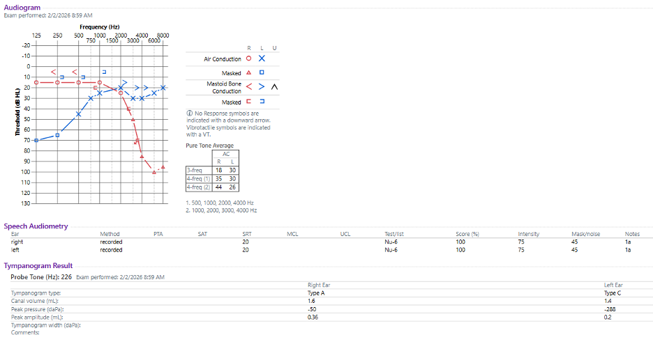
RIGHT ear: Type A; consistent with normal middle ear function, tympanic mobility, volume. ECV = 1.6 mL, peak pressure =-50 daPa, peak amplitude = 0.36 mL
LEFT ear: Type C; consistent with negative middle ear pressure. ECV = 1.4 mL, peak pressure = -288 daPa, peak amplitude = 0.2 mL
Speech Reception Threshold:
Right: 20 dB HL. Consistent with good SRT/PTA agreement.
Left: 20 dB HL. Consistent with good SRT/PTA agreement.
Word Recognition Score:
Right: 100 % @ 75 dB HL/45 dB masking (using Recorded NU-6 words) consistent with Excellent speech discrimination ability.
Left: 100 % @ 75 dB HL/45 dB masking (using Recorded NU-6 words) consistent with Excellent speech discrimination ability.
Pure tone audiometry:
Right: Normal hearing 125 through 2000 Hz and moderate to profound sensorineural hearing loss 3000 through 8000 Hz.
Left: Moderately-severe rising to mild conductive hearing loss 125 through 1000 Hz and normal hearing to borderline mild hearing loss 2000 through 8000 Hz.
This patient was recommended by the physician assistant (PA) to try a course of Fluticasone for presumed eustachian tube dysfunction, which he agreed to, but has not had follow up since. She also recommended following up with an otologist should his left ear problems continue unchanged or worsen. His ENT note with the PA indicates that he had interest in pursuing a hearing aid evaluation, which he did not complete with our facility.
Discussion
A fascinating aspect of this case is the possible protection of the left ear from sensorineural hearing loss due to the significant conductive component. This is hypothesized to be related to the reduced transfer of sound energy via air conduction (Park et al., 2016). The asymmetry is also consistent with his history as a musician, with the drum hi-hat, lead guitar, and horn section on his right side. It is also unknown whether his history of boxing contributed to his hearing loss, as he did not report any specific blow to one ear or the other that was worse than another. If we hypothesize that the sound level reaching the cochlea was reduced on the left compared to the right due to the middle ear conductive component functioning as ear protection, it may explain the asymmetry in his audiogram. It is also common for musicians to be more exposed on one side of their head due to their stage positions. There have been a handful of studies examining the possible protective effect of conductive hearing loss on noise exposure – primarily in industrial settings. More research is needed in this area.
References
- Forli, F., Capobianco, S., De Vito, A., Bruschini, L., & Lazzerini, F. (2025). Issues in the audiological assessment of otosclerosis. Acta Otorhinolaryngol Ital., Jun;45(Suppl. 1): S40-S48.
- Park, S.J., Sung, J.H., Sim, C.S., Yun, S.H., Yeom, J.H., Kwon, J-K., & Lee, J. (2016). Comparisons of hearing threshold changes in male workers with unilateral conductive hearing loss exposed to workplace noise: a retrospective cohort study for 8 years. Annals of Occupational and Environmental Medicine.28:51 DOI 10.1186/s40557-016-0132-1.
- Schachern, P.A., Paparella, M.M., Goycoolea, M.V., Duvall, A.J., & Choo, Y.B. (1987). The permeability of the round window membrane during otitis media, Arch Otolaryngol Head Neck Surg, Jun;113(6):625-9, doi: 10.1001/archotol.1987.01860060051014.
- Simpson, D.C., & O’Reilly, B.F. (1991). The protective effect of a conductive hearing loss in workers exposed to industrial noise. Clin Otolaryngol Allied Sci.Jun;16(3):274-7. doi: 10.1111/j.1365-2273.1991.tb00929.x. PMID: 1879071.
- Subramaniam, V., Ashkar, A., & Rai, S. (2020). Cochlear Dysfunction in Chronic Otitis Media and Its Determinants, Iranian Journal of Otorhinolaryngology, 32, 2, 79-84. doi: 10.22038/ijorl.2019.35045.2158
- Trevino, M., Zang, A., & Lobarinas, E. (2023). The middle ear muscle reflex: Current and future role in assessing noise-induced cochlear damage. The Journal of the Acoustical Society of America, 153(1), 436. https://doi.org/10.1121/10.0016853
- Walby, A.O., Barrera, A., & Schuknecht, H.F. (1983). Cochlear pathology in chronic suppurative otitis media, Ann Otol Rhinol Laryngol Suppl., Mar-Apr:103 Suppl:1-19.
- Zakrisson, J. E. (1975). The Role of the Stapedius Reflex in Poststimulatory Auditory Fatigue. Acta Oto-Laryngologica, 79(1–2), 1–10. https://doi.org/10.3109/00016487509124648