Close-Up Clinical Imaging of the Inner Ear. Coming To Audiology Soon?

From the Labs to the Clinics
Renowned auditory researcher Dr. Robert Harrison brings us up to date on information and research from the Labs. Appropriately titled “From the Labs to the Clinics”, Bob is involved in laboratory and applied/clinical research, including evoked potential and otoacoustic emission studies and behavioural studies of speech and language development in children with cochlear implants. For a little insight into Bob’s interests outside the lab and the clinic, we invite you to climb aboard Bob’s Garden Railway.
Creative Commons Attribution-Share Alike 3.0
CC BY-SA 3.0
Do you ever have patients with sensorineural hearing loss and wonder what the cochlea’s physical condition is? I’m sure that you do; I hope that you do! Perhaps you have an older person with presbycusis where you would like to see if the pattern of hair cell loss matches the audiogram. Perhaps a candidate for a cochlear implant where you wonder about spiral ganglion cell survival in the basal cochlear turn. Maybe a child with ANSD and a severe hearing loss where you would like to see the presence of outer hair cells responsible for a robust cochlear microphonic. You might even think to check if your patient with Meniere’s disease really does have endolymphatic hydrops.
To make these observations, we need a penetrating imaging technique with spatial resolution to show fine anatomical details. Standard CT or MRI do not come close. However, experimental work with high-energy X-rays generated by a synchrotron shows the potential for inner ear imaging. Such studies are being conducted by Canadian and US research teams that have recently published detailed X-ray images of the human cochlea. These figures reveal some of the fine structure of the cochlea that the new imaging technique can achieve. Structural details of the organ of Corti and the spiral ganglion are very clear. Even individual inner and outer hair cells can be resolved. The research team are based at Western University, Ontario, Harvard University, Boston, and a synchrotron facility in Saskatoon, Saskatchewan. (Janani et al., 2018).
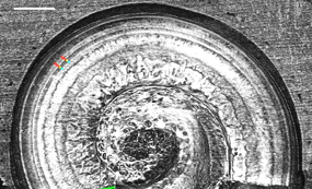
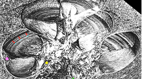
Synchrotron X-radiation is produced by charged particles traveling at extremely high speed by being forced to travel along curved paths by applied magnetic fields. To be clear, so far these images have only been obtained in cadaver temporal bone specimens, and not “in vivo.” So don’t try and order these X-rays quite yet!
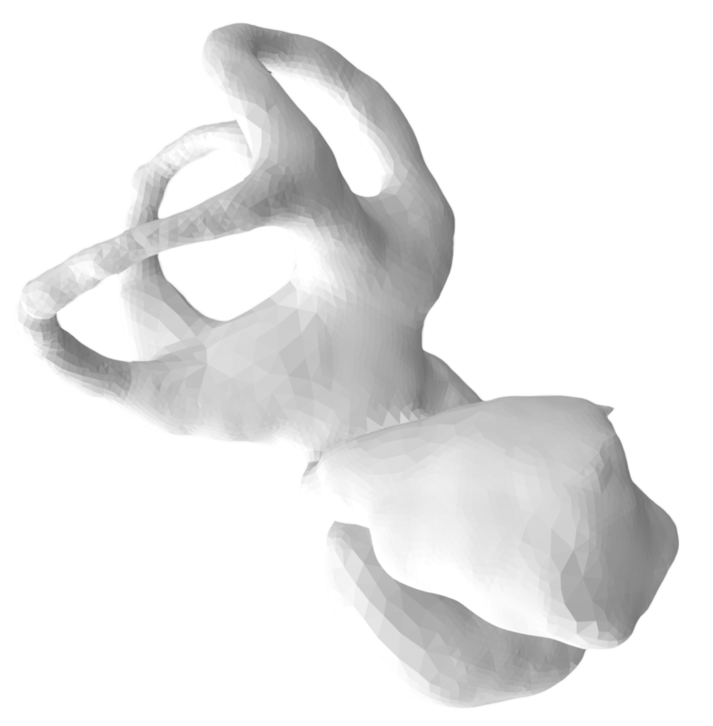
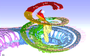
Such high-resolution X-ray data can be digitally enhanced and rendered into 3-D images where individual anatomical structures can be identified, highlighted, and quantified. Some examples of such 3-D visualization are shown below. These images are adapted from figures in Li et al., 2021.
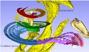
Whilst many technical aspects of synchrotron imaging of the inner ear have been explored, we are not at the point of having a clinical imaging tool. However, if this does become a reality, it could provide a very useful diagnostic tool in audiology.
References
- Iyer JS, Zhu N, Gasilov S, et al. Visualizing the 3-D cytoarchitecture of the human cochlea in an intact temporal bone using synchrotron radiation phase contrast imaging, Biomed. Opt. Express 2018;9, 3757-3767.
- Li, H., Helpard, L., Ekeroot, J. et al. Three-dimensional tonotopic mapping of the human cochlea based on synchrotron radiation phase-contrast imaging. Sci. Rep. 2021;11:4437. Available at: https://doi.org/10.1038/s41598-021-83225-w