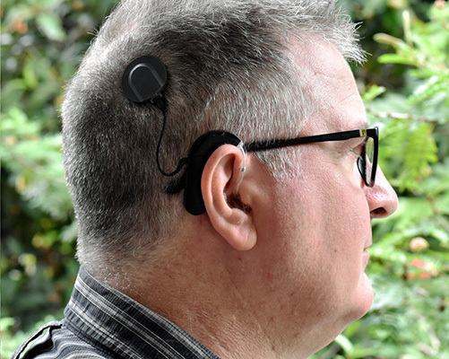
Vestibular Dysfunction Related to Cochlear Implantation in Adults

via Wikimedia Commons
Since the beginning of cochlear implant (CI) surgery, vertigo of vestibular origin has been described by many authors as a common post-operative side effect (Buchman et al., 2004; Krause et al., 2009). The cochlea and the vestibular end organs share common embryological origin, anatomical location, and similar physiology, this explains the high prevalence of vestibular symptoms peri-cochlear implantation. (Ibrahim et al., 2017; Swain et al., 2020; Vaz et al., 2022). According to Weinmann et al. (2021) CI recipients can be classified into 4 categories:
Group 0: No vertigo before and after surgery
Group 1: No vertigo before surgery, vertigo after surgery
Group 2: Vertigo before surgery, no vertigo after surgery
Group 3: Vertigo before and after surgery
A study done by Zawawi et al. (2014) involved 122 patients, in which dizziness was evident in 27% pre-CI and 45.9% post-CI. The commonest vestibular symptoms were unsteadiness followed by lightheadedness and immediate brief attacks of vertigo. The reported dizziness was mild in most of the patients.
Different mechanisms could lead to vestibular dysfunction during or after CI surgery. Including traumatic injury to the labyrinth during insertion of the electrode, perilymph loss during surgery, post-operative perilymphatic fistula, foreign body reaction with labyrinthitis, dislocation between 2 scalae leading to perilymphatic fluid leakage followed by fibrosis, endolymphatic hydrops. Moreover, the implant’s electrical stimulation of the labyrinth has been proposed as a mechanism to induce vertigo in CI recipients. In addition to that, benign paroxysmal positional vertigo has also been documented after CI. During the surgery, bone dust particles can enter the cochlea, through micro-injury or rupture of the basilar membrane. Afterwards, they move into the endolymphatic compartment of the scala media and block the endolymphatic pathway leading to hydrops. If these bone particles get into the semicircular canals, canalolithiasis and BPPV will develop. The vibratory damage from drilling to the inner ear can dislodge otoconia, another possible BPPV mechanism during CI surgery. (Katsiari et al., 2013, Ibrahim et al., 2017; Vaz et al., 2022).
Another theory suggests that dislodgment of an otolith by electrical stimulation can happen during initial fitting where the first fitting session may cause a triggering effect. (Katsiari et al., 2013; Ibrahim et al., 2017; Vaz et al., 2022). In 2020 researchers studied 101 CI recipients, they classified them into 4 groups: with early symptoms (n = 25), with late symptoms (n = 2), with pre-operative symptoms (n = 13), and with no symptoms (n = 61). Among the patients with early symptoms, 15 reported spontaneous vertigo attack, 6 reported only unsteadiness and/or lateropulsion, and 4 had other symptoms such as orthostatic vertigo, positional vertigo, visual tilt, and head-motion vertigo. These findings showed that 40% of the CI recipients develop vestibular complaints due to surgery or an already existing pre-operative condition. In the literature, 2 weeks after CI surgery are used to define early symptoms. Some CI recipients may experience dizziness several months or even a year after the surgery (Sosna-Duranowska et al., 2020). Most post-operative complaints were transient. Only a few cases had persistent dysfunction, usually in patients with existing inner ear pathologies and/or comorbidities. Several theories have been proposed to explain CI-associated vertigo. Most of them are due to post-operative ipsilateral vestibular dysfunction or due to BPPV. Pre-operative proper vestibular assessment is indicated to choose the accurate side for implant placement. For the post-operative, assessing symptomatic patients to provide appropriate management is necessary. Below is a list of practical assessment tools for conducting these assessments:
Dizziness Handicap Inventory questionnaire is 25 questions regarding the perceived severity of vertigo and its effects on quality of life. An 11-point difference is considered significantly different between repeated measures. (Jacobson & Newman ,1990; Rasmussen et al., 2021)
VOR and vestibular asymmetry assessment: Spontaneous nystagmus indicates vestibular asymmetry. The clinical head impulse test can identify an impaired vestibulo-ocular reflex in the tested ear, by observing the presence of a corrective saccade after a quick head movement. Moreover, head Shaking is a test of high-frequency vestibular asymmetry. Skull Vibration-induced nystagmus test (SVINT), nystagmus triggered by vibration applied to the skull. (Duams et al., 2011; Eggers et al., 2019).
Caloric testing using videonystgmography is a long-established measurement technique used in multiple studies to investigate lateral semicircular canal function after CI (Bittar et al., 2017). It has been frequently described that CI alters the caloric response in the implanted ear; however, controversial reports show no significant correlation between caloric test results and vertigo symptoms. Ibrahim et al. (2017) reported 39.5% pathologic cases preoperatively and 28% postoperatively.
Rotary chair testing examines the lateral semicircular canal’s function in the low and mid frequencies. However, its high cost, lack of specific canal information, and availability of video head impulse limit its usage.
Video Head Impulse Test. vHIT, according to Halmagyi and Curthoys is used to check the semicircular canals and the vestibulo-ocular reflex triggered by high-frequency head movements. (Halmagyi & Curthoys. 2018) The correlation between head and eye movements was registered as gain (= quotient eye speed ÷ head speed), with values below 0.8 considered abnormal. (Alfarghal M, et al. , 2022)
VEMP, the Cervical VEMP (cVEMP)which mainly reflects the function of the saccule and the ocular VEMP (oVEMP), which mainly reflects the function of the utricle (Li et al., 2020). In 80% to 100% of cases, cervical vestibular evoked myogenic potential (c VEMP) is usually lost, as per the metanalysis by Krause et al. (2009)
Postural test assessment (Romberg’s test). The patient is instructed to stand with their heels together and toes spread apart at approximately 30◦. The arms can rest along the body or be extended forward. The test is considered positive if the patient shifts or falls. The side of the fall will be the side of vestibular hypofunction. The test’s sensitivity can be increased if performed on a foam pad, first with the eyes open and then with the eyes closed. The test is positive when patients can stabilize their posture with their eyes open, but not when they are closed. The test on the pad simulates condition 5 of the dynamic posturography and the subject’s fall indicates vestibular dysfunction (Bittar et al., 2017).
Fukuda or Unterberger Test. With arms outstretched, the patient is asked to march in place 50 steps with their eyes closed. Rotations higher than 45° are considered abnormal. Asymmetrical lesions of the vestibular system result in body rotation toward the slow nystagmus component such as toward the hypofunctioning labyrinth (Grommes & Conway, 2011; Bittar et al., 2017).
Computerized Dynamic Posturography. An exam which is used as a quantitative assessment of the body balance. The postural performance of the CI patients significantly different from the controls, mainly in the Eyes Closed condition. The CI patients showed significantly reduced limits of stability and increased postural instability in static conditions. In dynamic conditions, they spent considerably more energy to maintain equilibrium. (Bernnard-Demanze et al., 2014; Ibrahim et al., 2020; Vaz et al., 2022)
Subjective virtual verticality. A straight line was drawn in the middle at the bottom of a bucket. The examiner held the bucket horizontally in front of the patient’s face. The patients could then look inside the bucket to see the line. The examiner spun the bucket 10 times in a row alternately to the left or right, and the patient should turn it back to the vertical using the line inside the bucket. The examiner then measured off a possible deviation from the vertical using a plumb and a protractor, which were attached to the bucket. SVV reflects lateral differences in the tonic affinity of the otolith organs (especially the utricle). A deviation of more than 2◦ was rated outside normal range (Böhmer et al., 1997).
Tests for BPPV: Dix-Hallpike and McClure-Paganini tests are used to diagnose BPPV.
Prevention
Insertion of electrode array through a round window rather than cochleostomy. This approach is the most advantageous and is still favoured in “structure-preserving surgery” (Todt et al., 2008). Slow electrode array insertion and topical application of corticosteroids intraoperatively help preserve vestibular function (Ibrahim et al., 2017; Vaz et al., 2022).
Treatment
Vestibular sedatives can be prescribed for reducing the acute onset vertigo after cochlear implantation, e.g., prochlorperazine, diphenhydramine and meclizine. Appropriate repositioning maneuver if BPPV is found. Vestibular rehabilitation is an effective intervention to improve overall balance, especially in cases of vestibular hypofunction. (Ibrahim et al., 2017; Vaz et al., 2022)
References
- Alfarghal M, Algarni MA, Sinha SK, Nagarajan A. VOR gain of lateral semicircular canal using video head impulse test in acute unilateral vestibular hypofunction: A systematic review. Front Neurol. 2022 Dec 8;13:948462. doi: 10.3389/fneur.2022.948462. PMID: 36570452; PMCID: PMC9773140.
- Bernard-Demanze L, Léonard J, Dumitrescu M, Meller R, Magnan J, Lacour M. Static and dynamic posture control in postlingual cochlear implanted patients: effects of dual-tasking, visual and auditory inputs suppression. Front Integr Neurosci. 2014 Jan 16;7:111. doi: 10.3389/fnint.2013.00111. PMID: 24474907; PMCID: PMC3893730
- Bittar RSM, Sato ES, Ribeiro DJS, Tsuji RK. Pre-operative vestibular assessment protocol of cochlear implant surgery: an analytical descriptive study. Braz J Otorhinolaryngol. 2017 Sep-Oct;83(5):530-535. doi: 10.1016/j.bjorl.2016.06.014. Epub 2016 Jul 31. PMID: 27574724; PMCID: PMC9444770
- Böhmer A. Zur beurteilung der otolithenfunktion mit der subjektiven visuellen vertikalen. HNO. (1997) 45:533–7. doi: 10.1007/s001060050127
- Buchman CA, Joy J, Hodges A, Telischi FF, Balkany TJ. Vestibular effects of cochlear implantation. Laryngoscope. (2004) 114(10 Pt 2 Suppl. 103):1–22.
- doi: 10.1097/00005537-200410001-00001
- Dumas G, Karkas A, Perrin P, Chahine K, Schmerber S. High-frequency skull vibration-induced nystagmus test in partial vestibular lesions. Otol Neurotol. 2011 Oct;32(8):1291-301. doi: 10.1097/MAO.0b013e31822f0b6b. PMID: 21897317.
- Eggers SDZ, Bisdorff A, von Brevern M, Zee DS, Kim JS, Perez-Fernandez N, Welgampola MS, Della Santina CC, Newman-Toker DE. Classification of vestibular signs and examination techniques: Nystagmus and nystagmus-like movements. J Vestib Res. 2019;29(2-3):57-87. doi: 10.3233/VES-190658. PMID: 31256095; PMCID: PMC9249296.
- Grommes C, Conway D. The stepping test: a step back in history. J Hist Neurosci. (2011) 20:29–33. doi: 10.1080/09647041003662255
- Katsiari E, Balatsouras DG, Sengas J, Riga M, Korres GS, Xenelis J. Influence of cochlear implantation on the vestibular function. Eur Arch Otorhinolaryngol. 2013;270(2):489–495. doi: 10.1007/s00405-012-1950-6.
- Krause E, Louza JPR, Wechtenbruch J, Hempel J-M, Rader T, Gürkov R.Incidence and quality of vertigo symptoms after cochlear implantation.J Laryngol Otol. (2009) 123:278–82. doi: 10.1017/S002221510800 296X
- Halmagyi, G & Curthoys, Ian & Halmagyi, Michael. (2018). The video head impulse test in clinical practice. Neurological Sciences an
- Jacobson GP, Newman CW. The development of the dizziness handicap inventory. Arch Otolaryngol Head Neck Surg. (1990) 116:424–7. doi: 10.1001/archotol.1990.01870040046011
- Ibrahim I, da Silva SD, Segal B, Zeitouni A. Effect of cochlear implant surgery on vestibular function: meta-analysis study. J Otolaryngol Head Neck Surg. 2017 Jun 8;46(1):44. doi: 10.1186/s40463-017-0224-0. PMID: 28595652; PMCID: PMC5465585.
- Li X, Gong S. The Effect of Cochlear Implantation on Vestibular Evoked Myogenic Potential in Children. Laryngoscope. 2020 Dec;130(12):E918-E925. doi: 10.1002/lary.28520. Epub 2020 Feb 7. PMID: 32031698; PMCID: PMC7754474.
- Rasmussen KMB, West N, Tian L, Cayé-Thomasen P. Long-Term Vestibular Outcomes in Cochlear Implant Recipients. Front Neurol. 2021 Aug 11;12:686681. doi: 10.3389/fneur.2021.686681. PMID: 34456848; PMCID: PMC8385200.
- Sosna-Duranowska M, Tacikowska G, Gos E, Krupa A, Skarzynski PH, Skarzynski H. Vestibular Function After Cochlear Implantation in Partial Deafness Treatment. Front Neurol. 2021 May 21;12:667055. doi: 10.3389/fneur.2021.667055. PMID: 34093414; PMCID: PMC8175845.
- Swain SK, Achary S, Das SR. Vertigo in pediatric age: Often challenge to clinicians. Int J Cur Res Rev. 2020;12(18):136-41.
- Todt I, Basta D, Ernst A. Does the surgical approach in cochlear implantation influence the occurrence of post-operative vertigo? Otolaryngol Head Neck
- Surg. (2008) 138:8–12. doi: 10.1016/j.otohns.2007.09.003
- Weinmann C, Baumann U, Leinung M, Stöver T, Helbig S. Vertigo Associated With Cochlear Implant Surgery: Correlation With Vertigo Diagnostic Result, Electrode Carrier, and Insertion Angle. Front Neurol. 2021 Jun 11; 12:663386. doi: 10.3389/fneur.2021.663386. PMID: 34177768; PMCID: PMC8226011.
- Vaz FC, Petrus L, Martins WR, Silva IMC, Lima JAO, Santos NMDS, Turri-Silva N, Bahmad F Jr. The effect of cochlear implant surgery on vestibular function in adults: A meta-analysis study. Front Neurol. 2022 Aug 10;13:947589. doi: 10.3389/fneur.2022.947589. PMID: 36034277; PMCID: PMC9402268.
- Zawawi F, Alobaid F, Leroux T, Zeitouni AG. Patients reported outcome post-cochlear implantation: how severe is their dizziness? J Otolaryngol Head Neck Surg. 2014 Dec 10;43(1):49. doi: 10.1186/s40463-014-0049-z. PMID: 25492268; PMCID: PMC4273446

